Note: Your progress in watching these videos WILL NOT be tracked. These training videos are the same videos you will experience when you take the full ProPALS program. You may begin the training for free at any time to start officially tracking your progress toward your certificate of completion.
There can be many forms of bradycardia. Commonly seen blocks include sinus bradycardia, and for multiple blockages, complete and 3rd-degree heart block.
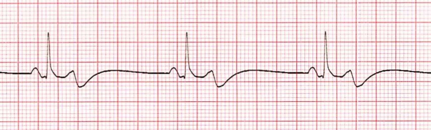
In this lesson, we'll look more closely at an example of what bradycardia looks like on an ECG for a pediatric patient and see what findings and measurements lead us to that conclusion.
It's vital to remember that if there are signs of bradycardia, regardless of whatever underlying reasons that are causing the patient to display symptoms related to bradycardia, we must first treat for the bradycardia, as it takes precedent over those underlying causes.
 *Bradycardia ECG
1. The Heart Rhythm
The first thing you'll want to look at is the heart rhythm. Does the heart rhythm look regular? Or does it look irregular? In the above graphic, it's regular.
2. The Heart Rate
Next, you'll want to look at the heart rate of the patient. What is the patient's heart rate? Is it normal? Or is it too slow or too fast? In this case, it's too slow, as the rate is less than 60 beats per minute.
Remember, to determine the patient's heart rate you'll want to observe the following areas on the ECG paper printout and perform the following calculations.
The horizontal axis of ECG paper grids is where time is measured. Each small square is 1mm in length and represents .04 seconds. Each larger square is 5mm in length and represents .2 seconds. Therefore a 6-second interval would be 30 large squares.
To determine the heart rate, count the number of QRS complexes over this 6-second interval and multiply by 10.
It's also important to understand that when it comes to pediatric patients, normal heart rates vary based on the age of the patient. For example, a normal heart rate for a 12-year-old will probably be bradycardic for an infant or a newborn.
3. P-Wave
After looking at the heart rate, check to see if the patient's P-waves look normal by asking yourself the following few questions.
- Are the patient's P-waves present? In this case, the answer is yes.
- Do they occur regularly? The answer is yes again.
- Is there one P-wave for each QRS complex? Yes, there is.
- Are the P-waves smooth, rounded, and upright? The answer is again yes.
- Do all the P-waves have a similar shape? Yes, they all have a similar shape.
4. PR Interval
Next, look at the PR interval on the patient's ECG readout and ask yourself the following questions:
- Is the PR interval normal, meaning less than .20 seconds or is it contained within one large square on the readout? The answer is yes, it's less than .20 seconds and contained within one large square.
- Is the PR interval constant? Yes, it is.
5. QRS Complex
The last thing you should look at to determine if the sinus rhythm is normal or not is the QRS complex and ask yourself these questions while you do:
- Is the QRS interval less than .09 seconds? Yes, it is.
Remember, as long as the QRS fits within two small squares on the ECG printout and is not greater than two and one-quarter small squares, it's within the normal range.
- Is the QRS complex wide or narrow? In this case, it's narrow.
- Are the QRS complexes similar in appearance or are there noticeable differences? In this case, we can see that each looks similar.
So, what is your cardiac interpretation? Based on these questions and on the findings from the ECG readout above, it's safe to say that this patient is in sinus bradycardia.
- We have a regular rhythm.
- We have a slower than normal heart rate, at less than 60 beats per minute.
- The P-waves look normal, with each being followed by a QRS complex.
- The PR interval is less than .20 seconds.
- The QRS is less than .09 seconds.
Proper oxygenation and ventilation are crucial to raise the heart rate for a child with this ECG bradycardic readout.
Pro Tip: For a child in bradycardia with a pulse rate less than 60 and signs of poor perfusion, despite oxygen and ventilation, you must begin chest compressions followed by full CPR immediately.
Additional Bradycardia Information
Bradycardia is defined as a heart rate that is slow when compared with a normal heart rate range for that specific child's age, his or her level of activity, and his or her clinical condition.
Symptomatic Bradycardia
Symptomatic bradycardia is defined as a heart rate slower than normal for the child's age, which is usually less than 60 beats per minute, associated with cardiopulmonary compromise.
Cardiopulmonary Compromise
Cardiopulmonary compromise is defined as hypotension, acutely altered mental status, as in a decreased level of consciousness, and signs of shock.
Warning: Bradycardia is an unfortunate sign of impending cardiac arrest in all pediatric patients, particularly when it is associated with hypotension and/or symptoms of poor tissue perfusion. Which is why we advise immediate CPR, in spite of adequate oxygenation and ventilation, for all pediatric patients whose heart rate is less than 60 beats per minute.


